Refractive Lense Exchange Toronto – No Bifocals
Refractive Lens Exchange replaces the crystalline lens of the eye with an intraocular lens. This procedure is recommended over laser eye surgery when patients desire…
Refractive Lens Exchange replaces the crystalline lens of the eye with an intraocular lens. This procedure is recommended over laser eye surgery when patients desire…

Family traditions are a beautiful thing. Perhaps your own family has special recipes for certain holidays or particular vacation spots you return to over and…

Serena Ryder, Canada’s celebrity singer-songwriter, Juno award winner (2008, 2009, 2010) underwent successful vision correction surgery by Dr Raymond Stein at the Bochner Eye Institute.

We are delighted and proud of our 90th year of ophthalmology at the Bochner Eye Institute, a unique milestone in North America. The Bochner Eye…
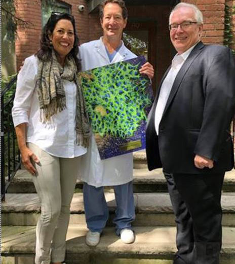
Dr. Raymond Stein, the medical director of Bochner Eye Institute, was recently honored for serving 10 years on the Board of Directors of Fighting Blindness…

Everyone’s eyes occasionally feel gritty or uncomfortable. But for some people, the dry, scratchy sensation is an ongoing problem. Dry eye syndrome occurs when the…

Millions of people live free from the hassle of glasses or contact lenses, thanks to LASIK. If you have worn visual aids for most of…

According to recent research papers, cataract surgery has more benefits than we previously thought. Not only does the surgery make patients’ lives better with clearer…

Cataracts are a clouding of the eye’s natural lens and one of the leading causes of vision loss in Americans. The normal aging process is…

One of the most common questions Bochner Eye Institute receives from men and women considering LASIK is, How long does the procedure take? Many people…